Consultant Vascular & Endovascular Surgeon • Hyderabad
Endovascular or open repair planned from aneurysm size, growth, symptoms, anatomy, life expectancy and procedural fitness.
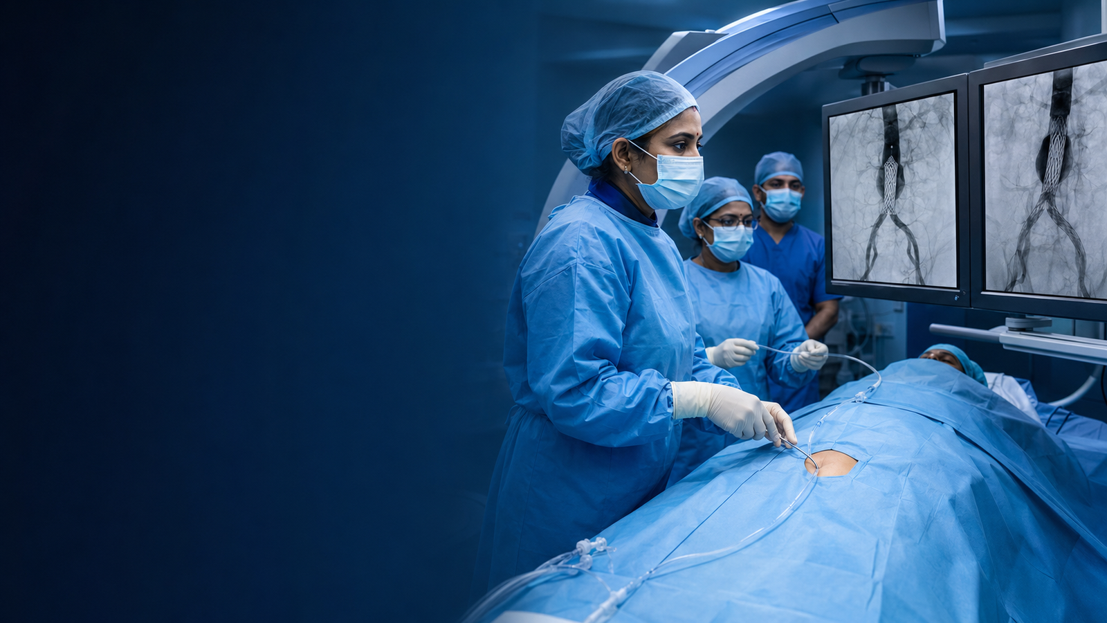
Aortic aneurysm repair prevents rupture by excluding or replacing the weakened segment. Endovascular aneurysm repair, or EVAR, places a stent graft through arteries in the groin. Open repair replaces the diseased aorta through a larger operation. EVAR generally involves smaller incisions and faster early recovery, while open repair may offer advantages for selected anatomy and requires a different long-term surveillance pathway.
EVAR advances a stent graft through groin arteries to seal the aneurysm
Open repair clamps the aorta and replaces the weak segment with a graft
Blood flow to branch arteries is protected according to aneurysm location
Post-repair imaging checks the graft and aneurysm sac
Bring every prior aneurysm scan so growth can be confirmed. Stop tobacco, optimise blood pressure and follow individual instructions for antiplatelets, anticoagulants, diabetes medicines, fasting and kidney protection. Dental, skin or urinary infections should be reported.
Open repair requires major anaesthesia and intensive perioperative planning. EVAR may use general, regional or local techniques depending on anatomy, device and patient health.
EVAR often has a shorter initial recovery than open repair, but both require activity, wound and medicine instructions. Open recovery commonly takes several weeks. Exact timing depends on aneurysm complexity and health before surgery.
Both approaches carry risks of bleeding, heart or lung complications, kidney injury, bowel or limb circulation problems, infection and death. EVAR can develop endoleak, device movement or later reintervention; open repair has greater early physiological stress.
Aneurysms below a repair threshold are commonly monitored with scheduled imaging while cardiovascular risks are treated. Repair may not offer net benefit when operative risk or limited life expectancy outweighs rupture prevention.
EVAR requires lifelong imaging to check the seal, device and aneurysm sac. Open repair also needs clinical follow-up and selective imaging. Sudden severe abdominal or back pain, fainting or collapse requires emergency care even after prior repair.

Bring original scans, written reports, current medicines, previous procedure notes and a short timeline of symptoms.
These questions cover suitability, preparation, procedure, recovery, risks, alternatives and follow-up.
Aortic aneurysm repair prevents rupture by excluding or replacing the weakened segment. Endovascular aneurysm repair, or EVAR, places a stent graft through arteries in the groin. Open repair replaces the diseased aorta through a larger operation. EVAR generally involves smaller incisions and faster early recovery, while open repair may offer advantages for selected anatomy and requires a different long-term surveillance pathway.
It may be considered for people with an aneurysm that has reached an appropriate repair threshold, rapid growth or symptoms attributable to the aneurysm, anatomy suitable for a durable endovascular or open repair, overall health and life expectancy that support preventive treatment. A consultation and imaging are required because a matching symptom does not automatically make someone a procedure candidate.
Aortic Aneurysm Repair may be minimally invasive, open surgical or a coordinated combination depending on the exact technique. The incision, hospital stay and physiological stress should be explained for the option actually being considered.
Assessment may include thin-slice ct angiography and measurement review, comparison with earlier scans for growth, heart, lung, kidney and functional assessment, discussion of evar anatomy, open repair and surveillance. Additional tests are selected according to age, kidney function, other illnesses and the urgency of treatment.
The planned steps can include evar advances a stent graft through groin arteries to seal the aneurysm, open repair clamps the aorta and replaces the weak segment with a graft, blood flow to branch arteries is protected according to aneurysm location, post-repair imaging checks the graft and aneurysm sac. The treating team should explain the exact device or surgical approach, alternatives and the reason it fits the patient's anatomy.
Open repair requires major anaesthesia and intensive perioperative planning. EVAR may use general, regional or local techniques depending on anatomy, device and patient health.
EVAR often has a shorter initial recovery than open repair, but both require activity, wound and medicine instructions. Open recovery commonly takes several weeks. Exact timing depends on aneurysm complexity and health before surgery.
Both approaches carry risks of bleeding, heart or lung complications, kidney injury, bowel or limb circulation problems, infection and death. EVAR can develop endoleak, device movement or later reintervention; open repair has greater early physiological stress.
Aneurysms below a repair threshold are commonly monitored with scheduled imaging while cardiovascular risks are treated. Repair may not offer net benefit when operative risk or limited life expectancy outweighs rupture prevention.
EVAR requires lifelong imaging to check the seal, device and aneurysm sac. Open repair also needs clinical follow-up and selective imaging. Sudden severe abdominal or back pain, fainting or collapse requires emergency care even after prior repair.
A structured vascular review can help you understand suitability, alternatives and next steps.
Specialist evaluation and treatment for conditions affecting arteries, veins, circulation, wounds and dialysis access.
Clinic contact details and consultation timings can be added after confirmation.
© 2026 Dr. Pragna Vascular. Privacy Policy · Medical Disclaimer