Consultant Vascular & Endovascular Surgeon • Hyderabad
DVT and Venous Interventions
Anticoagulation-led care with catheter-based clot treatment or venous reconstruction for carefully selected patients.
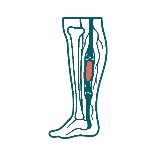
What Is DVT and Venous Interventions?
Most deep vein thrombosis is treated with anticoagulation, which helps prevent clot extension and pulmonary embolism while the body gradually processes the clot. Catheter-directed thrombolysis, mechanical thrombectomy, angioplasty or venous stenting may be considered in selected patients with extensive acute clot, threatened venous circulation or significant chronic obstruction. These procedures are not routine for every calf DVT.
Who May Be Considered?
- Extensive acute iliofemoral DVT with severe symptoms
- A limb threatened by severe venous obstruction
- Selected chronic iliac or central venous obstruction
- Acceptable bleeding risk and anatomy likely to benefit
Assessment and Tests
- Venous Doppler and clot-location review
- CT or MR venography when pelvic veins are involved
- Bleeding risk, kidney function and medicine review
- Symptom duration and post-thrombotic impact assessment
How Treatment May Be Performed
Enter the affected vein through a small access site
Use medicine, mechanical devices or both to reduce selected clot burden
Treat a significant underlying venous narrowing when appropriate
Continue anticoagulation and compression according to the final diagnosis
How to Prepare
Report every blood thinner, recent operation, stroke, bleeding episode, pregnancy possibility, kidney problem and contrast allergy. Do not delay emergency evaluation for breathlessness or chest pain while arranging an elective consultation.
Comfort and Anaesthesia Planning
Venous interventions commonly use local anaesthesia and sedation. Procedures involving prolonged thrombolytic infusion may require monitored hospital care, and the plan changes with clot extent and patient condition.
What to Expect During Recovery
Observation ranges from several hours to a longer admission. Bruising at the access site and residual swelling can occur. Anticoagulation usually continues, and compression may be advised. Improvement can be gradual, especially when a vein has been obstructed for a long time.
Risks and Limitations
Bleeding is the central risk of clot-dissolving treatment and can rarely be severe. Other risks include access-site bleeding, contrast or kidney problems, vessel injury, pulmonary embolism, incomplete clot removal, re-thrombosis and stent-related issues.
Alternatives to Consider
Anticoagulation alone is the standard pathway for many patients. Compression, exercise and long-term symptom management may be appropriate for post-thrombotic disease. An IVC filter has limited indications and is not a substitute for anticoagulation when blood thinners can be used.
Follow-Up After Treatment
Medication adherence, bleeding review and repeat imaging are planned individually. Recurrent swelling, new chest symptoms or bleeding on anticoagulation needs prompt assessment. Long-term follow-up evaluates post-thrombotic symptoms and venous-stent patency where applicable.
Discuss Treatment With Dr. Pragna
Bring original scans, written reports, current medicines, previous procedure notes and a short timeline of symptoms.
Frequently Asked Questions About DVT and Venous Interventions
These questions cover suitability, preparation, procedure, recovery, risks, alternatives and follow-up.
Most deep vein thrombosis is treated with anticoagulation, which helps prevent clot extension and pulmonary embolism while the body gradually processes the clot. Catheter-directed thrombolysis, mechanical thrombectomy, angioplasty or venous stenting may be considered in selected patients with extensive acute clot, threatened venous circulation or significant chronic obstruction. These procedures are not routine for every calf DVT.
It may be considered for people with extensive acute iliofemoral dvt with severe symptoms, a limb threatened by severe venous obstruction, selected chronic iliac or central venous obstruction, acceptable bleeding risk and anatomy likely to benefit. A consultation and imaging are required because a matching symptom does not automatically make someone a procedure candidate.
DVT and Venous Interventions may be minimally invasive, open surgical or a coordinated combination depending on the exact technique. The incision, hospital stay and physiological stress should be explained for the option actually being considered.
Assessment may include venous doppler and clot-location review, ct or mr venography when pelvic veins are involved, bleeding risk, kidney function and medicine review, symptom duration and post-thrombotic impact assessment. Additional tests are selected according to age, kidney function, other illnesses and the urgency of treatment.
The planned steps can include enter the affected vein through a small access site, use medicine, mechanical devices or both to reduce selected clot burden, treat a significant underlying venous narrowing when appropriate, continue anticoagulation and compression according to the final diagnosis. The treating team should explain the exact device or surgical approach, alternatives and the reason it fits the patient's anatomy.
Venous interventions commonly use local anaesthesia and sedation. Procedures involving prolonged thrombolytic infusion may require monitored hospital care, and the plan changes with clot extent and patient condition.
Observation ranges from several hours to a longer admission. Bruising at the access site and residual swelling can occur. Anticoagulation usually continues, and compression may be advised. Improvement can be gradual, especially when a vein has been obstructed for a long time.
Bleeding is the central risk of clot-dissolving treatment and can rarely be severe. Other risks include access-site bleeding, contrast or kidney problems, vessel injury, pulmonary embolism, incomplete clot removal, re-thrombosis and stent-related issues.
Anticoagulation alone is the standard pathway for many patients. Compression, exercise and long-term symptom management may be appropriate for post-thrombotic disease. An IVC filter has limited indications and is not a substitute for anticoagulation when blood thinners can be used.
Medication adherence, bleeding review and repeat imaging are planned individually. Recurrent swelling, new chest symptoms or bleeding on anticoagulation needs prompt assessment. Long-term follow-up evaluates post-thrombotic symptoms and venous-stent patency where applicable.
Bring Your Scans and Current Treatment Advice
A structured vascular review can help you understand suitability, alternatives and next steps.

Specialist evaluation and treatment for conditions affecting arteries, veins, circulation, wounds and dialysis access.
Vascular Care
Patient Information
Appointments
Clinic contact details and consultation timings can be added after confirmation.
© 2026 Dr. Pragna Vascular. Privacy Policy · Medical Disclaimer