Consultant Vascular & Endovascular Surgeon • Hyderabad
Embolisation and Sclerotherapy
Image-guided treatment for selected vascular malformations after precise classification of vessel type, flow and tissue involvement.
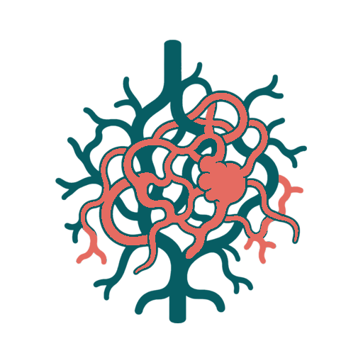
What Is Embolisation and Sclerotherapy?
Embolisation blocks abnormal vessels from inside using catheters and specialised materials, while sclerotherapy injects a medicine that irritates and closes selected abnormal vascular spaces. These techniques are used for particular vascular malformations, pelvic venous disorders and other vascular problems. Treatment is planned from imaging and flow classification because a fast-flow arteriovenous malformation is managed differently from a slow-flow venous malformation.
Who May Be Considered?
- Pain, bleeding, swelling, ulceration or functional limitation
- A lesion with imaging-defined vessels that can be targeted
- Pelvic venous reflux or obstruction with matching symptoms
- A staged treatment goal agreed by the multidisciplinary team
Assessment and Tests
- Clinical examination and symptom correlation
- Doppler to assess vessel type and flow
- MRI, CT venography or angiography according to the lesion
- Review of skin, nerve, muscle, bone and organ involvement
How Treatment May Be Performed
Use image guidance to enter the target vessel or malformation
Deliver an embolic or sclerosant chosen for the vessel pattern
Protect normal circulation and nearby tissue
Stage treatment when complete treatment in one sitting would increase risk
How to Prepare
Report pregnancy possibility, kidney disease, allergies, blood thinners and prior treatment materials. Bring original MRI, CT and angiography images. Fasting, admission, anaesthesia and medicine instructions vary substantially with the lesion.
Comfort and Anaesthesia Planning
Small treatments may use local anaesthesia and sedation, while painful, extensive or high-flow procedures may require general anaesthesia. The expected tissue reaction and procedure length guide the plan.
What to Expect During Recovery
Pain, swelling and bruising can occur as treated vessels react. Observation and return to activity depend on site and extent. Several staged sessions may be required, and symptom improvement can take time as swelling settles.
Risks and Limitations
Risks include skin or tissue injury, ulceration, nerve damage, bleeding, clot migration, infection, allergic or contrast reaction, non-target embolisation and recurrence. Fast-flow malformations can have major haemodynamic and bleeding risks.
Alternatives to Consider
Observation, compression, pain management, surgery or combined treatment may be suitable. Some malformations are safer to monitor than to treat. A scan abnormality alone is not enough reason for embolisation when symptoms do not correlate.
Follow-Up After Treatment
Clinical review and repeat imaging evaluate response and plan any next stage. Treatment usually aims to control symptoms and complications rather than promise permanent eradication of every abnormal channel.
Discuss Treatment With Dr. Pragna
Bring original scans, written reports, current medicines, previous procedure notes and a short timeline of symptoms.
Frequently Asked Questions About Embolisation and Sclerotherapy
These questions cover suitability, preparation, procedure, recovery, risks, alternatives and follow-up.
Embolisation blocks abnormal vessels from inside using catheters and specialised materials, while sclerotherapy injects a medicine that irritates and closes selected abnormal vascular spaces. These techniques are used for particular vascular malformations, pelvic venous disorders and other vascular problems. Treatment is planned from imaging and flow classification because a fast-flow arteriovenous malformation is managed differently from a slow-flow venous malformation.
It may be considered for people with pain, bleeding, swelling, ulceration or functional limitation, a lesion with imaging-defined vessels that can be targeted, pelvic venous reflux or obstruction with matching symptoms, a staged treatment goal agreed by the multidisciplinary team. A consultation and imaging are required because a matching symptom does not automatically make someone a procedure candidate.
Embolisation and Sclerotherapy may be minimally invasive, open surgical or a coordinated combination depending on the exact technique. The incision, hospital stay and physiological stress should be explained for the option actually being considered.
Assessment may include clinical examination and symptom correlation, doppler to assess vessel type and flow, mri, ct venography or angiography according to the lesion, review of skin, nerve, muscle, bone and organ involvement. Additional tests are selected according to age, kidney function, other illnesses and the urgency of treatment.
The planned steps can include use image guidance to enter the target vessel or malformation, deliver an embolic or sclerosant chosen for the vessel pattern, protect normal circulation and nearby tissue, stage treatment when complete treatment in one sitting would increase risk. The treating team should explain the exact device or surgical approach, alternatives and the reason it fits the patient's anatomy.
Small treatments may use local anaesthesia and sedation, while painful, extensive or high-flow procedures may require general anaesthesia. The expected tissue reaction and procedure length guide the plan.
Pain, swelling and bruising can occur as treated vessels react. Observation and return to activity depend on site and extent. Several staged sessions may be required, and symptom improvement can take time as swelling settles.
Risks include skin or tissue injury, ulceration, nerve damage, bleeding, clot migration, infection, allergic or contrast reaction, non-target embolisation and recurrence. Fast-flow malformations can have major haemodynamic and bleeding risks.
Observation, compression, pain management, surgery or combined treatment may be suitable. Some malformations are safer to monitor than to treat. A scan abnormality alone is not enough reason for embolisation when symptoms do not correlate.
Clinical review and repeat imaging evaluate response and plan any next stage. Treatment usually aims to control symptoms and complications rather than promise permanent eradication of every abnormal channel.
Bring Your Scans and Current Treatment Advice
A structured vascular review can help you understand suitability, alternatives and next steps.
Specialist evaluation and treatment for conditions affecting arteries, veins, circulation, wounds and dialysis access.
Vascular Care
Patient Information
Appointments
Clinic contact details and consultation timings can be added after confirmation.
© 2026 Dr. Pragna Vascular. Privacy Policy · Medical Disclaimer